Menu Navigation

Services
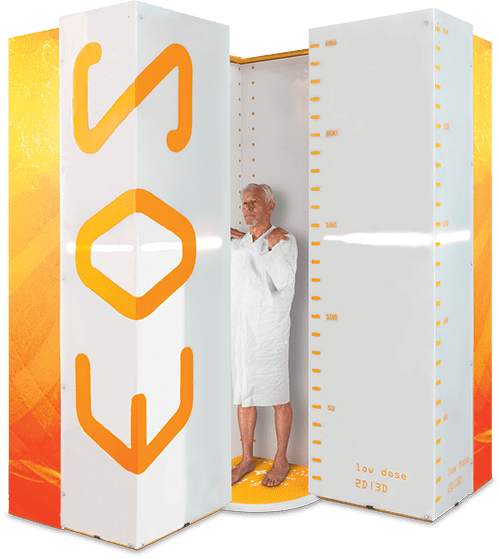
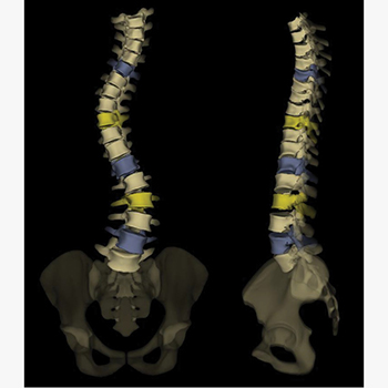
The EOS system delivers low dose stereoradiographic images of patients in functional position. The unique biplanar design and vertical scanning allows the acquisition of full body with precise 2D and 3D measurements, helping clinicians to better visualize mechanisms between the spine, hip, and knee, enhancing patient treatment across all stages of care.
The EOS system introduces a fifth modality to reduce patient radiation dose for a wide range of patients providing an exceptional image contra
The EOS system can reduce a patient’s radiation dose by 95% when substituted for specific CT exams. EOS exams reduce radiation dose by 50% compared to DR systems and 85% compared to CR systems.
High-accuracy imaging data from EOS allows providers to plan treatment strategies around each patient’s unique 3D anatomy. Our low dose imaging system helps medical experts and their patients co-create the best path to wellness.
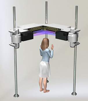
During an EOS exam, the patient stands or sits in an upright position inside a special scanning cabin. Two very narrow X-ray beams – one vertical, one horizontal – scan the entire body to create 2D and 3D images of the spine and joints. Unlike traditional X-ray imaging, where the patient may have to be repositioned to get views from different angles, these two simultaneous scans provide all the imaging necessary. Capturing frontal and lateral (side-view), full-body images take less than twenty seconds. If a full-body image is not necessary (such as for a knee condition), the EOS system can be set to scan a particular region of the patient’s anatomy

EOS exams are unique because they capture the front and side view of your body at the same time. These images are of consistent high quality from head to toe - with no magnification. That means accurate diagnoses, measurements and 3D models without high radiation or costly, time-consuming exams. With EOS, your medical team can offer personalized care based on your unique anatomy throughout your care journey.
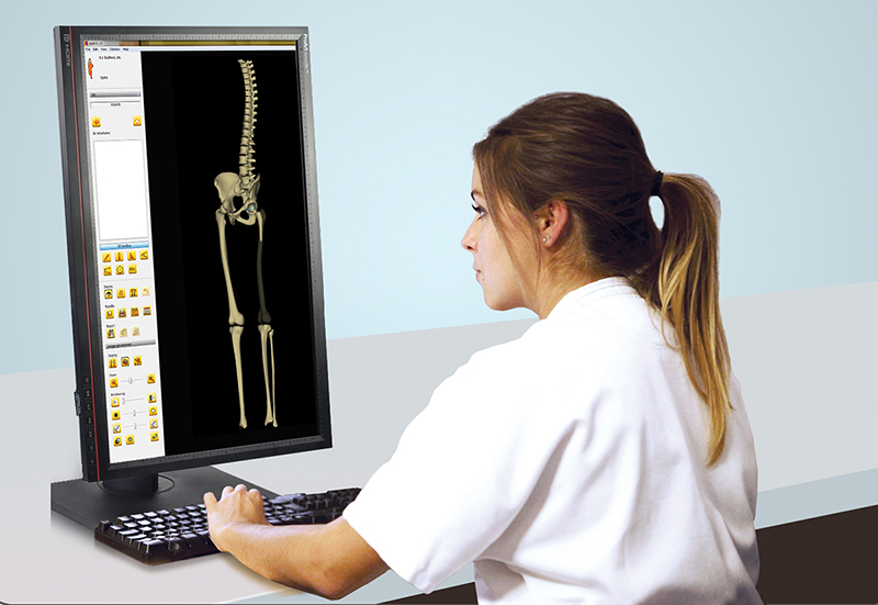
The images from your patient’s EOS imaging exam are available immediately. Because EOS creates 3-D images, there is no need to piece together multiple images. Our radiologist and orthopaedic specialists will review the results and discuss the results with patients’ family and determine next steps.

